Glossary

What is X-Ray?

X-Ray is a quick, painless imaging test that uses electromagnetic waves to create pictures of the inside of the body, especially bones. X-Rays help doctors and chiropractors see fractures, infections, arthritis. Or spinal misalignments without surgery. The test takes only a few minutes and exposes patients to a small amount of radiation.

Reviewed by ChiropractorBrentwood.comSources reviewed: U.S. Food and Drug Administration - Radiation-Emitting Products: X-Rays, American Chiropractic Association - Diagnostic Imaging

Quick Facts About X-Ray

Category

Medical imaging

Used for

Bone fractures, spinal alignment, joint health

Common confusion

X-Rays show bones, not soft tissues like muscles or ligaments

Also called

Radiograph, Röntgenogram

Often discussed with

Car Accident Injury Treatment, Work Injury Treatment

Key Takeaways About X-Ray

Understanding X-Ray

X-Ray in Chiropractor: X-Ray is a quick, painless imaging test that uses electromagnetic waves to—visual guide

X-Ray is a type of medical imaging that uses a controlled beam of electromagnetic radiation to create pictures of structures inside the body. When the beam passes through the body, dense materials like bones absorb more radiation and appear white on the image. While softer tissues appear in shades of gray. This contrast helps healthcare providers identify problems such as broken bones, infections. Or abnormal growths without invasive procedures.

Related glossary terms: Chiropractic Adjustment, Joint Dysfunction, Herniated Disc.

X-Rays are widely used in chiropractic care because they provide clear views of the spine and joints. A chiropractor may order an X-Ray to check for spinal misalignments, degenerative changes like arthritis. Or other conditions that could affect treatment plans. The images help ensure that adjustments and therapies are safe and custom to the patient’s specific needs. While X-Rays are not needed for every patient, they're valuable when symptoms suggest underlying structural issues.

How X-Ray Works?

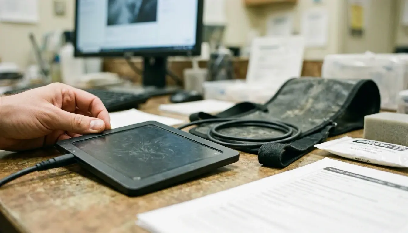
An X-Ray machine sends a focused beam of radiation through the part of the body being examined. A detector on the other side captures the radiation that passes through, creating an image based on how much radiation different tissues absorb. Bones, which contain calcium, absorb the most radiation and appear white on the image. Soft tissues, like muscles and organs, absorb less and appear in darker shades of gray. Air, such as in the lungs, appears black because it absorbs almost no radiation.

For local customers, The entire process typically takes less than five minutes. Patients are positioned between the X-Ray machine and the detector. And they may be asked to hold still or take a deep breath to improve image clarity. Protective lead aprons or shields are often used to cover areas of the body not being imaged to cut down on radiation exposure. The resulting images are reviewed by a radiologist or chiropractor to identify any abnormalities or guide treatment decisions.

Why X-Ray Matters?

How X-Ray applies to Chiropractor services in Brentwood, United States—practical illustration

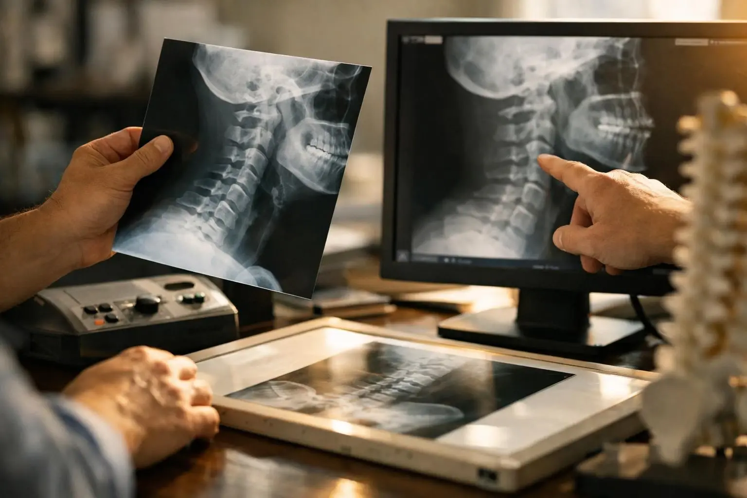
X-Rays play a critical role in diagnosing and treating musculoskeletal conditions. For patients experiencing pain, limited mobility. Or injury, an X-Ray can reveal the root cause, such as a fracture, dislocation. Or degenerative disease. This information allows chiropractors and other healthcare providers to develop targeted treatment plans that address the specific issue rather than relying on guesswork. For example, an X-Ray might show a herniated disc or spinal misalignment, guiding the chiropractor to use gentle adjustments or other therapies to relieve pressure on nerves.

Beyond diagnosis, X-Rays also help monitor progress over time. Follow-up images can show whether a fracture is healing correctly, if arthritis is worsening. Or if a treatment plan is effective. This ongoing assessment ensures that patients receive the most appropriate care and can make adjustments as needed. While X-Rays involve some radiation exposure, the benefits of accurate diagnosis and treatment often outweigh the risks, especially when used judiciously.

When X-Ray Matters Most?

X-Rays are most useful in situations where structural issues are suspected but not visible from the outside. For example, if a patient has persistent back pain after an injury, an X-Ray can determine whether a fracture or spinal misalignment is contributing to the discomfort. Similarly, X-Rays are commonly used after car accidents or falls to check for hidden injuries, such as hairline fractures or joint dislocations, that might not cause immediate symptoms but could lead to long-term problems if left untreated.

X-Rays are also valuable for patients with chronic conditions like arthritis or scoliosis. Regular imaging can track the progression of these conditions and help providers adjust treatment plans to slow deterioration or manage symptoms. And X-Rays are often used before certain chiropractic procedures, such as spinal adjustments, to ensure the treatment is safe and appropriate for the patient’s anatomy. By providing a clear view of the spine and joints, X-Rays help chiropractors avoid potential complications and tailor their approach to each patient’s unique needs.

How to Evaluate X-Ray?

Related Concepts Compared

X-Ray vs. MRI

MRI uses magnetic fields and radio waves to create detailed images of soft tissues like muscles, ligaments. And organs. While X-Rays focus on bones and some dense structures.

X-Ray vs. CT Scan

A CT scan combines multiple X-Ray images taken from different angles to create cross-sectional views of the body, offering more detail than a single X-Ray.

Expert Note

X-Rays are a foundational tool in musculoskeletal care. But they should be used thoughtfully. Not every patient needs an X-Ray. And repeated exposure should be minimized, especially for children and pregnant women. Always discuss the necessity and benefits with your provider.

Common Mistakes or Myths About X-Ray

  • Assuming X-Rays show soft tissues like muscles or ligaments—they primarily show bones.
  • Believing X-Rays are always necessary for chiropractic care—many treatments can proceed without them.
  • Thinking X-Rays are dangerous due to radiation—modern X-Rays use very low doses and are generally safe when used appropriately.

X-Ray in Practice: A Real-World Example

After a car accident, a patient visits a chiropractor complaining of neck pain. The chiropractor orders an X-Ray to check for whiplash-related injuries. The X-Ray reveals a slight misalignment in the cervical spine, allowing the chiropractor to perform gentle adjustments to realign the vertebrae and relieve the patient’s pain.

Sources & Further Reading on X-Ray

Related Services

Related Terms

Chiropractic Adjustment

Chiropractic Adjustment is a hands-on procedure where a licensed chiropractor applies controlled force to a specific joint, usually in the spine, to improve alignment, reduce pain. And restore movement. The goal is to correct joint dysfunction without surgery or medication, relying on the body’s natural ability to heal.

Joint Dysfunction

Joint Dysfunction is a condition where a joint in the body fails to move or function normally, often causing pain, stiffness. Or reduced range of motion. It can result from injury, overuse, poor posture. Or underlying conditions like arthritis. Joint Dysfunction may affect any joint, including the spine, shoulders, hips. Or knees. And can disrupt daily activities if left untreated.

Herniated Disc

Herniated Disc is a spinal condition where the soft, gel-like center of a spinal disc pushes through a tear in the disc’s tougher outer layer. This bulging or ruptured material can press on nearby nerves, causing pain, numbness. Or weakness in the back, neck, arms. Or legs. Herniated discs often occur due to aging, injury.

Spinal Decompression

Spinal Decompression is a non-surgical treatment designed to relieve pressure on the spine and discs. It involves gently stretching the spine using a motorized table or traction device to create negative pressure within the discs, which may help herniated or bulging discs retract and promote healing by allowing oxygen, water. And nutrients to flow into the affected area.

Orthopedic Tests

Orthopedic Tests are hands-on examinations performed by healthcare providers to assess musculoskeletal conditions, such as joint injuries, nerve compression. Or muscle strains. These tests involve specific movements, pressure. Or positioning to reproduce symptoms and identify the source of pain or dysfunction. They help guide diagnosis and treatment without relying solely on imaging or lab results.

ChiropractorBrentwood.com

Have Questions About X-Ray?

Contact ChiropractorBrentwood.com for practical guidance on X-Ray and related chiropractor work in Brentwood.

+1 615-777-0624