Understanding Electromyography

Electromyography, often called EMG, is a diagnostic tool used to assess the health of muscles and the nerves that control them. When muscles contract, they produce tiny electrical signals. Electromyography captures these signals using electrodes, which are small sensors placed on the skin or inserted into the muscle. The test helps doctors determine if muscle weakness, pain. Or numbness is caused by a problem in the muscle itself or in the nerves supplying it.
Electromyography is commonly used alongside other tests, such as nerve conduction studies, to provide a complete picture of nerve and muscle function. While the idea of electrical activity in muscles might sound complex, the test itself is straightforward and non-invasive for most patients. The electrodes simply record the natural electrical activity that occurs when muscles move, making it a safe and effective way to gather important diagnostic information.
How Electromyography Works?
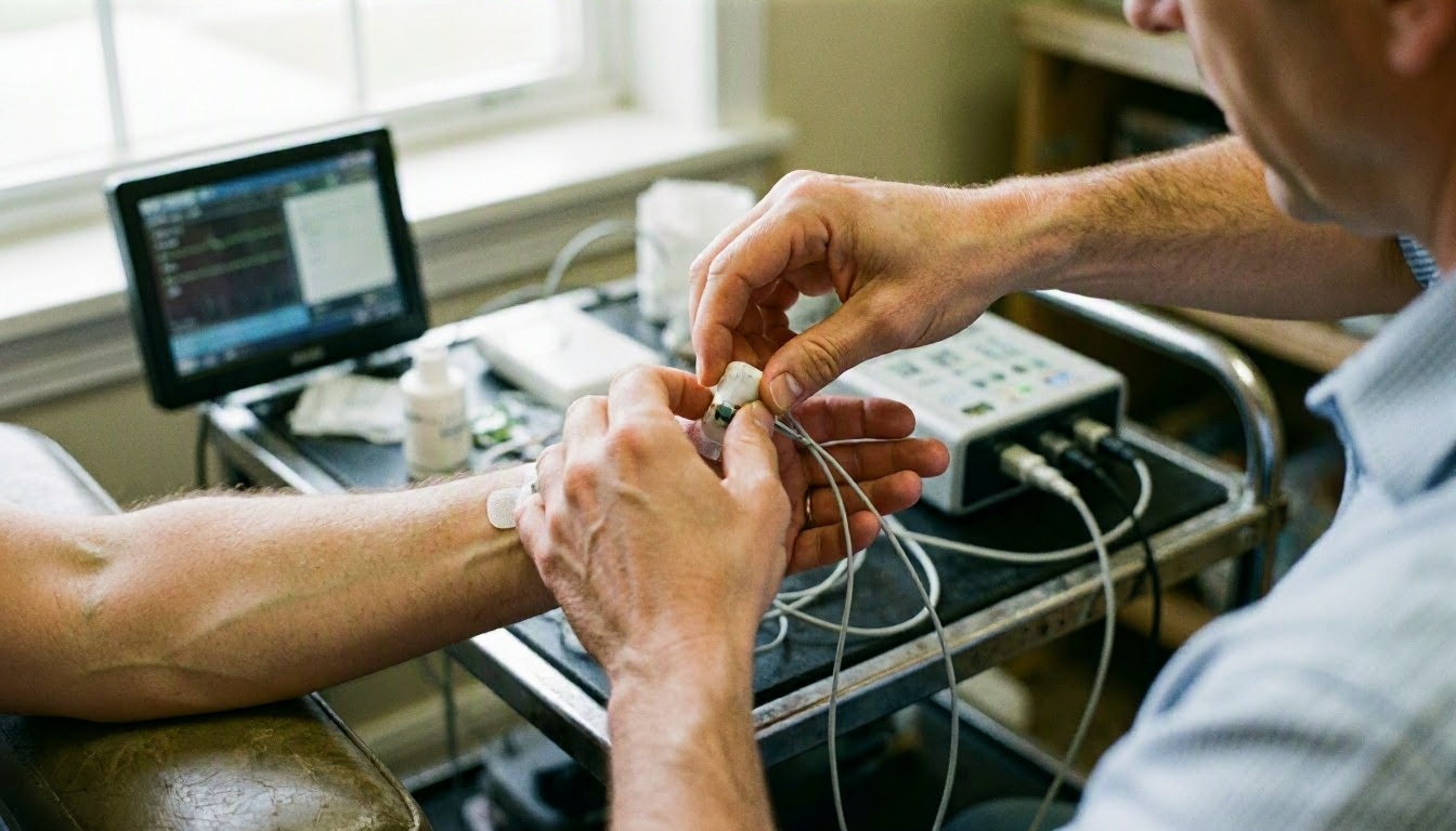
During an electromyography test, a healthcare provider places electrodes on the skin over the muscle being tested. In some cases, a thin needle electrode is inserted directly into the muscle to get a more detailed reading. The patient is asked to contract the muscle, such as bending an arm or flexing a foot. While the electrodes record the electrical activity. The signals are displayed on a monitor as waveforms, which the provider analyzes to detect abnormalities.
The test typically takes 30 to 60 minutes, depending on how many muscles are being examined. While the needle insertion may cause slight discomfort, most patients tolerate the procedure well. The results are interpreted by a specialist, such as a neurologist or physiatrist, who looks for patterns that indicate nerve damage, muscle disease. Or other conditions. For example, abnormal electrical activity at rest may suggest a muscle disorder. While reduced activity during contraction could indicate nerve damage.
Why Electromyography Matters?
Electromyography plays a crucial role in diagnosing conditions that cause muscle weakness, pain. Or numbness. Without this test, it can be difficult to determine whether symptoms are due to a muscle problem, nerve damage. Or another underlying issue. For instance, electromyography can help distinguish between sciatica, which involves nerve compression. And a muscle strain, which affects the muscle fibers directly. This distinction is important because the treatments for these conditions differ significantly.
The test also helps guide treatment decisions by providing objective data about muscle and nerve function. For example, if electromyography reveals nerve damage, a doctor might recommend physical therapy, medication. Or in some cases, surgery to relieve pressure on the nerve. Conversely, if the test shows muscle disease, the treatment plan may focus on strengthening exercises or other interventions to improve muscle function. By pinpointing the source of symptoms, electromyography helps ensure patients receive the most appropriate care for their condition.
When Electromyography Matters Most?
Electromyography is most useful when patients experience unexplained muscle weakness, pain, tingling. Or numbness. These symptoms can arise from a variety of causes, including nerve compression, muscle diseases. Or injuries. For example, someone with carpal tunnel syndrome may feel numbness or tingling in their hand. While a person with a herniated disc in the spine might experience sciatica, which causes pain radiating down the leg. In these cases, electromyography can confirm whether the symptoms are due to nerve damage or another issue.
A practical next step is The test is also valuable for monitoring the progression of certain conditions or assessing the effectiveness of treatment. For instance, if a patient undergoes surgery to relieve nerve compression, follow-up electromyography can show whether the nerve function is improving. And electromyography is often used in research to study muscle and nerve diseases, helping scientists develop better treatments. Whether for diagnosis, treatment planning. Or monitoring, electromyography provides critical insights that can improve patient outcomes.